I have degree in chemical engineering (BSc; METU, Turkey), biotechnology (PhD; Cranfield University, UK) and more than 14 years professional experience in the area of biosensors. After completing a master’s degree in biochemistry (MSc; GYTE, Turkey), I worked at two University of Cambridge spin-off companies (Affinity Sensors and Akubio Ltd.) at different positions. Some of the experiences I obtained during my professional work and PhD study include sensor chip fabrication, sensor surface chemistry development, biological assay development (for the detection of proteins, DNA, virus and bacteria), sensor signal enhancement via nanoparticles and product development from idea to market. Not only did I use different commercial biosensor instruments (optical, piezoelectric and electrochemical) but was also involved in the development of new biosensor devices and sensor chips from prototype to commercial products, and some of the products I developed or was involved in its development are on the market.
Biosensor technology is an exciting area that involves many disciplines. From physics to understand how the instruments work, to the knowledge of immunoassays, protein-protein interaction, DNA hybridisation, viral and bacterial detection, it involves many interesting areas. One of the most valuable contributions of biosensors is that they allow detection of clinically relevant molecules, so they are very important diagnostic tools of our day.
I have been working at BILGEM-TUBITAK (The Scientific and Technological Research Council of Turkey) since August 2011 as a Senior Researcher. Since 2014, I have been working as a Department Manager and also lead the Bioelectronics Devices and Systems project team. BILGEM, as an institute of TUBITAK (The Scientific and Technological Research Council of Turkey), has one of the best infrastructures in Turkey, and performs cutting-edge research and product development particularly on optoelectronic and microelectronic areas. The microelectronics team has capability of 0.7µm CMOS IC production within 10 class clean room, the optoelectronics team manufactures e.g. CMOS digital smart area cameras and multi-purpose CCD line cameras and markets to the industry. BILGEM institute also specialises in software solutions for security that has great importance for secure data transfer between a biosensor device and remote data analysis centers. In addition to that Bioinformatics team specialises in biologic data analysis solutions. Therefore the team of BILGEM is very experienced in electronics, optics and software areas that are must for a state of the art biosensor device design.
My research interests include:
Molecular recognition, sensing and diagnostics
Nanotechnology applications in analytical and life sciences
Genetic and protein biomarker selection and detection
Biosensor assay development and signal enhancement
Cancer, as one of the leading causes of death in the world, is caused by malignant cell division and growth that depends on rapid DNA replication. To develop anti-cancer drugs this feature of cancer could be exploited by utilizing DNA-damaging molecules. To achieve this, the paraben substituted cyclotetraphosphazene compounds have been synthesized for the first time and their effect on DNA has been investigated. For the first time, a new screening method has been used to investigate the DNA damage, which is based on an automated biosensor device that relies on the real-time electrochemical profiling (REPTM) technology.
Although electroanalytical techniques/biosensors have been proven to be a useful tool for genotoxicity tests, lack of automated and fast devices prevented its use as the routine toxicology analysis tools. In a review paper published by Rusling et al. in 2007, it had been stated that if the sensing devices had been inexpensive and better developed, they had potential to be used for toxicity screening for drug development. To answer the need, here the use of a new automated biosensor device (MiSens) has been described, where the assay has been performed using a microfluidic channel integrated biochip with the sequential injections of the assay reagents. MiSens biosensor device relies on the Real-time Electrochemical Profiling (REPTM) technology and can be utilized to investigate the genotoxicity by measuring the DNA hybridization efficiency on the biochip surface. The DNA hybridization (that is the formation of the DNA duplex by annealing two complementary single strands) is achieved by implementing a surface immobilized capture probe and a hybridized target and detection probes.
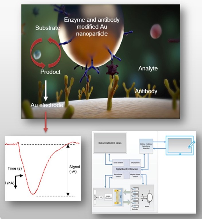
Figure 1. MiSens biosensor and the biochip.
It is believed that the data obtained from the biosensor assays help researchers to choose the most probable drug candidates out of many, as this method directly and quantitatively shows the effect of the compounds to the DNA strand. Hence, this pre-screening method can be applied prior to the conventional in vitro and in vivo genotoxicity tests.
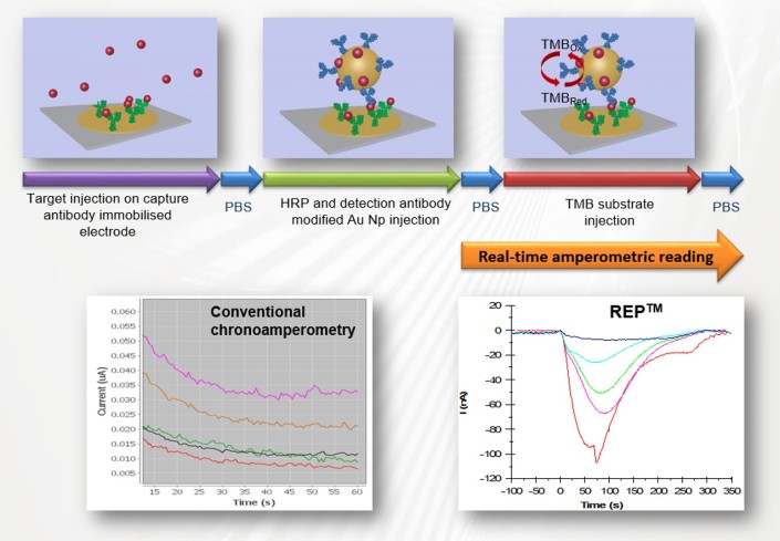
Figure2. Schematics of the DNA damage test using MiSens device and the biochip.
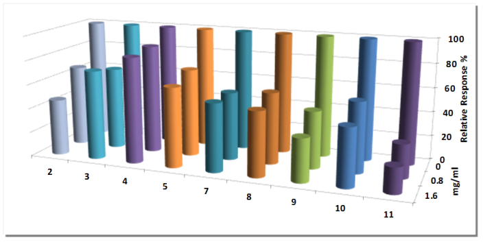
Fig. 3 The amperometric responses obtained from the REP assay have been shown in the figure as percent relative responses. Varying concentrations of the compounds (0, 0.8 and 1.6 mg/mL) have been incubated with target and detection DNA probes prior to the injection on to the biochip.
Most of the biosensor publications describe only the results for proof of principle of the analyte detection using electrochemical biosensors and the number of studies performed using integrated systems with the necessary fluidics is quite limited. In addition mostly, the assay procedures are long, labourous and not suitable for automation. In response to that, we have developed a new detection technology, which we term Real-time Electrochemical Profiling (REPTM).
While this technology relies on the fundamental basics of electrochemical immunosensing, in particular amperometry, it has several key features including a new electrode array, microfluidics based assay and real-time amperometric measurements during the flow of enzyme substrate. For the common flow injection electrochemical immunoassay platforms, initially in a separate container or fluidic system the enzyme immunoassay is performed, and later the reaction solution is sent to (usually) a bare electrode for the detection at a certain flow rate. However with the REP system, we were able to combine both steps together on one electrode surface. That distinguishes our sensing platform from other flow injection based electrochemical immunoassays. In addition, whole assay is performed during the reagents flow; therefore, there is no need to stop the flow either during the analyte binding or enzymatic reaction, hence the sensing can be achieved reasonably fast.
A new fully automated and microfluidics integrated biosensor MiSens has been developed that utilises REP technology for sensing.
The smallest microfluidics integrated and fully automated electrochemical biosensor: MiSens
Here we describe a new integrated and fully automated lab-on-a-chip based biosensor device ‘MiSens’. The key features of the MiSens include a new electrode array, an integrated microfluidic system and real-time amperometric measurements during the flow of enzyme substrate.
For the device, a new plug and play type sensor chip docking station has been designed that with one move it enables the formation of a ~ 7 µl capacity flow cell on the electrode array with the necessary microfluidic and electronic connections. The MiSens device has been developed by our multi-disciplinary team by integrating and automatising the earlier developed sensing platform REPTM (Real-time Electrochemical Profiling).
While simple protocols such as prime and desorb can be controlled from the LCD display on the device, other main device control procedures can be run wireless by a tablet using the MiContTM software developed by the team. MiContTM interface enables user to create a list of assay steps that forms the test protocol. This protocol can be saved and re-used when needed. During data acquisiton the software shows the electrochemical measurements real time.
BILGEM, as an institute of TUBITAK (The Scientific and Technological Research Council of Turkey), has one of the best infrastructures in Turkey, and performs cutting-edge research and product development particularly on optoelectronic and microelectronic areas. BILGEM has a highly advanced and well equipped “Biomedical Electronics Devices and Systems Development Group” which has been established as a consequence of bringing together its competencies in opto-electronics and software engineering with its expertise in chemistry-biology-nanotechnology.
In various fields like medicine, environmental control, food security and military defense; there is an increasing demand for on-site detection, fast identification and urgent response which brings the necessity to employ laboratory detection procedures on standalone automatic devices. In response to that TUBITAK BILGEM’s Bioelectronic Devices and Systems Group has been developing portable and fully automated biosensor devices using optical and electrochemical biosensor detection techniques.
Developed systems and the devices
MiSens™; is a mobile, fully automated biosensor device, that can be used in vehicles or on a desk. Can be controlled from the remote control app on a tablet PC or its’ on-device touch screen with minimal training of the user. Includes easy-to- use biochips (Patent Application No. PCT/IB2015/052479).
BiSens™; is a handheld, on-site biosensor equipment, which allows the detection of analytes without any necessity of an expert user or a laboratory owing to its single use cartridge system.
SOBE™; is an optical, handheld, transcutaneous bilirubin measurement device which helps in the diagnosis of jaundice seen in the newborn babies (design patent application No. 2015/01029).
Fast and sensitive detection of mycotoxins in wheat using novel electrode arrays and Real-time Electrochemical Profiling
In the literature several studies can be found related with mycotoxin detection using electrochemical techniques; however, most of these publications describe the results of the tests that are performed using bench top potentiostats with off the shelf electrodes and require – hands on work. This shows that most of the publications describe only the results for proof of principle of the toxin detection using biosensors and the number of studies performed using integrated systems with the necessary fluidics is quite limited. In addition mostly, the assay procedures are long, labourous and not suitable for automation. In response to that, we have developed a novel detection technology, which we term Real-time Electrochemical Profiling (REPTM). While this technology relies on the fundamental basics of electrochemical immunosensing, in particular amperometry, it has several key features including a novel electrode array (Patent no: PT 2014/39932), microfluidics based assay and real-time amperometric measurements during the flow of enzyme substrate. The proposed REP platform consists of new electrode arrays that are easy to fabricate, has a small imprint allowing microfluidic system integration, enables multiplexed amperometric measurements and performs well in terms of electrochemical immunoassay detection as shown through the deoxynivalenol detection assays.
The deoxynivalenol detection has been conducted according to an optimised REP assay protocol using deoxynivalenol standards at varying concentrations and a standard curve was obtained (y = -20.33ln(x)+124.06; R2 = 0.97) with an LOD of 6.25 ng/ml. As both ELISA and REP detection methods use HRP as the label and TMB as the substrate; the performance of the REP platform as an ELISA reader also has been investigated and a perfect correlation between the DON concentration and the current response was obtained (y=-14,56ln(x)+101.02; R2 = 0.99). The calibration curves of both assays have been compared to conventional ELISA tests for confirmation. After assay optimization using toxin spiked buffer, the DON detection assay has also been performed to detect toxins in wheat grain.
(A) Raw data of the REPTM assay for DON detection, where the amperometric measurement was taken in real time during the injection of TMB substrate (50 µL/min). The obtained current is proportional to the amount of the surface bound DON-HRP conjugate and inversely proportional to the DON concentration in sample. (B) Linear and (C) logarithmic calibration curves for the DON detection assay.
More information can be found from our recent poster: Esen, Z. Olcer, T. Muhammad, A. Ersoy, S. Budak, Y. Uludag, “Fast and sensitive detection of mycotoxins with Real-time Electrochemical Profiling and nanoparticles amplification system“,6th Lab-On-A-Chip European Congress, 10-11.03.2014, Berlin, Germany.
The Effects of Electrode Design to The Electrochemical Biosensor Performance
In the current study, a novel electrode array and integrated microfluidics have been designed and characterised in order to create a sensor chip that is not only easy, rapid and cheaper to produce but also have a smaller imprint and good electrochemical sensing properties. The current study includes the assessment of the effects of an Au quasi-reference electrode and the use of shared reference/counter electrodes for the array, in order to obtain a small array that could be produced using a fine metal mask. In the study, it was found that when Au is used as quasi-reference electrode, the arrays with shared reference and counter electrodes results in faster electron transfer kinetics and prevents the potential change with respect to scan rate, and hence advantageous with respect to conventional electrodes. In addition, the resulting novel electrode array, has been shown to result in higher current density (10.52 µA/cm2; HRP detection assay) and measured diffusion coefficient (14.40×10⁻¹² cm2/s; calculated from the data of cyclic voltammetry with 1 mM potassium ferricyanide) with respect to the conventional electrodes tested in the study. Using the new electrode arrays, the detection limits obtained from horse radish peroxidase (HRP) and bisphenol A assays were 12.5 ng/ml (2.84×10⁻¹⁰ M ) and 10 ng/ml (44×10−9 M), respectively. Performing the HRP detection assay in a flow injection system using arrayintegrated microfluidics, provided 25 times lower detection limit (11.36×10⁻¹² M), although Ti has been used as electrode material instead of Au. In short, incorporation of this new electrode array to lab-on-a-chip or MEMs (micro-electro mechanic systems) technologies may pave the way for easy to use, automated biosensing devices that could be used for a variety of applications from diagnostics to environmental monitoring, and studies will continue to move forward in this direction.
More information can be found from our recent poster displayed at Biosensing Technologies 2013 conference in Spain:
Reference:
Uludag, Y.; Olcer, Z.; Sagiroglu, M. S., Design and Characterisation of a Thin-film Electrode Array with Shared Reference/Counter Electrodes for Electrochemical Detection. Biosensors and Bioelectronics 2014, DOI:10.1016/j.bios.2014.01.048.
In most cases biomarker detection tests using label-free biosensors were performed only in buffered pure solutions rather than serum. To obtain clinically relevant results, it is essential to perform the biomarker test in human serum. The main difficulty of using serum as the assay media is high non-specific interaction between the sensor surface and serum proteins. A number of strategies have been employed to reduce the non-specific binding of clinical samples; such as:
use of mixed self-assembled monolayer coatings which contains ethylene glycol units
carboxy methyl dextran surface
addition of additives to assay buffer
use of blocking agents after antibody immobilisation such as milk or certain polymers
use of serum as running buffer to eliminate the mismatch between the running buffer and sample containing serum
diluting the serum till effects of non-specific binding is minimised
The above described methods can either be applied individually or together to reduce the non-specific binding of serum proteins achieving different success. Each one of them has different efficiency and may have some drawbacks. In our study we have designed a new buffer (we call it matrix elimination buffer, in short martix buffer) that eliminates 98% of serum protein non-specific binding and enables assays using high concentrations of human serum. You can find more information from our recent poster displayed at Biosensors 2010 conference in Glasgow:
and from our recently published paper:
Y. Uludag, I.E. Tothill, “Development of a Sensitive Detection method for Cancer Biomarkers in Human Serum (75%) using a Quartz Crystal Microbalance Sensor and Nanoparticles amplification system”, Talanta, Vol 82(1), 277-282. PDF
Nucleic acid based recognition of viral sequences can be used together with label-free biosensors to provide rapid, accurate confirmation of viral infection. To enhance detection sensitivity, gold nanoparticles can be employed with mass-sensitive acoustic biosensors (such as a quartz crystal microbalance) by either hybridising nanoparticle-oligonucleotide conjugates to complimentary surface-immobilised ssDNA probes on the sensor, or by using biotin-tagged target oligonucleotides bound to avidin-modified nanoparticles on the sensor. We have evaluated and refined these signal amplification assays for the detection from specific DNA sequences of Herpes Simplex Virus (HSV) type 1 and defined detection limits with a 16.5 MHz fundamental frequency thickness shear mode acoustic biosensor.
Results
In the study the performance of semi-homogeneous and homogeneous assay formats (suited to rapid, single step tests) were evaluated utilising different diameter gold nanoparticles at varying DNA concentrations. Mathematical models were built to understand the effects of mass transport in the flow cell, the binding kinetics of targets to nanoparticles in solution, the packing geometries of targets on the nanoparticle, the packing of nanoparticles on the sensor surface and the effect of surface shear stiffness on the response of the acoustic sensor. This lead to the selection of optimised 15 nm nanoparticles that could be used with a 6 minute total assay time to achieve a limit of detection sensitivity of 5.2 × 10⁻¹² M. Larger diameter nanoparticles gave poorer limits of detection than smaller particles. The limit of detection was three orders of magnitude lower than that observed using a hybridisation assay without nanoparticle signal amplification.
Conclusions
An analytical model was developed to determine optimal nanoparticle diameter, concentration and probe density, which allowed efficient and rapid optimisation of assay parameters. Numerical analysis and subsequent associated experimental data suggests that the response of the mass sensitive biosensor system used in conjunction with captured particles was affected by i) the coupled mass of the particle, ii) the proximal contact area between the particle and the sensor surface and iii) the available capture area on the particle and binding dynamics to this capture area. The latter two effects had more impact on the detection limit of the system than any potential enhancement due to added mass from a larger nanoparticle.


 MiSens™; is a mobile, fully automated biosensor device, that can be used in vehicles or on a desk. Can be controlled from the remote control app on a tablet PC or its’ on-device touch screen with minimal training of the user. Includes easy-to- use biochips (Patent Application No. PCT/IB2015/052479).
 ng a novel electrode array (Patent no: PT 2014/39932), microfluidics based assay and real-time amperometric measurements during the flow of enzyme substrate. The proposed REP platform consists of new electrode arrays that are easy to fabricate, has a small imprint allowing microfluidic system integration, enables multiplexed amperometric measurements and performs well in terms of electrochemical immunoassay detection as shown through the deoxynivalenol detection assays.


